
Categories Uncategorized

Why Some Blood Tests Need to Be Repeated

Why Did My Doctor Order a Repeat Blood Test? Understanding the Reasons

Leaving a clinic only to get a follow-up call requesting a second blood draw can feel unsettling. It is completely natural for your mind to jump to worst-case scenarios, but the reality is far more routine: repeating a blood test is a standard clinical practice designed to ensure accuracy, safety, and proper diagnosis.

A single blood sample provides a snapshot of a single moment in time. Understanding why your healthcare provider needs a second look can help turn health anxiety into clarity.

1. Biological Fluctuations and Time-of-Day Variations

The human body is dynamic, and biomarker levels shift naturally throughout the day:

  • Hormonal rhythms: Hormones like cortisol and testosterone peak early in the morning and decline by the afternoon.
  • Hydration and diet: Dehydration can falsely elevate kidney markers like creatinine or alter electrolyte concentrations.
  • Temporary stress or illness: Mild infections, strenuous workouts, or high physical stress can temporarily spike white blood cell counts or liver enzymes without signaling a chronic issue.

A repeat test helps establish your true baseline rather than capturing an isolated spike or dip.

2. Verifying Borderline or Anomalous Results

Diagnostic reference ranges are built around statistical averages across large populations. When a biomarker lands just outside the standard cutoff—such as slightly elevated fasting glucose or marginally low thyroid-stimulating hormone (TSH)—doctors rarely diagnose on that single data point.

Re-testing allows your doctor to confirm whether the anomaly persists or naturally settles back into normal ranges before prescribing medication or ordering invasive procedures.

3. Strict Fasting or Preparation Protocols

Certain blood panels require strict pre-test preparation that can easily go off track:

  • Lipid panels and fasting glucose: Require 8 to 12 hours of complete fasting (water only).
  • Medication timing: Specific therapeutic drug levels must be drawn right before your next scheduled dose (trough level).
  • Supplements: High-dose biotin (vitamin B7) can significantly interfere with immunoassays for cardiac troponin and thyroid hormones.

If your initial sample was taken without meeting these conditions, a repeat draw under controlled guidelines is essential for valid numbers.

4. Technical and Laboratory Factors

Even with automated, state-of-the-art laboratory standards, pre-analytical sample issues occur:

IssueWhat It MeansWhy It Requires a Redraw
HemolysisRed blood cells rupture during collection or transitArtificially elevates potassium and alters liver enzymes
Clotted SampleMicro-clots form in anticoagulated tubesInvalidates complete blood count (CBC) and platelet counts
Insufficient Volume (QNS)“Quantity Not Sufficient” for all ordered panelsThe lab cannot run every necessary assay

5. Monitoring Treatment Response and Disease Progression

Repeat testing is also an intentional therapeutic tool. For chronic condition management, scheduled repeat tests evaluate whether an intervention is working effectively:

  • HbA1c: Checked every 3 to 6 months to assess long-term blood sugar control.
  • Thyroid panels: Repeated 6 to 8 weeks after adjusting levothyroxine dosages.
  • Inflammatory markers (CRP, ESR): Tracked to evaluate how autoimmune conditions or infections respond to therapy.

How to Prepare for Your Repeat Blood Draw

To make sure your second test goes smoothly:

  1. Clarify instructions: Confirm exact fasting requirements and whether regular morning medications should be taken or held until after the draw.
  2. Hydrate well: Drink plenty of plain water to make veins easier to access and keep plasma volume steady.
  3. Rest before collection: Avoid intense exercise within 24 hours of tests checking muscle enzymes (CK), liver markers, or kidney function.

Getting called back for a repeat blood test is rarely a medical emergency—it is simply your medical team prioritizing diagnostic precision over guesswork.
Categories Uncategorized

What a Lipid Profile Actually Tests

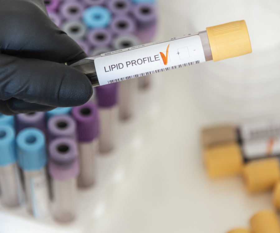
Getting your blood drawn for a routine checkup often ends with a lab report full of medical jargon: LDL, HDL, triglycerides, VLDL, total cholesterol. While it is easy to glance at the numbers and wait for your doctor to say “looks good,” understanding what a lipid profile actually measures puts your cardiovascular health directly into your hands.

A lipid panel is a simple blood test that measures the fats (lipids) circulating in your bloodstream. These molecules are essential for building cell membranes and producing hormones, but when they fall out of balance, they can increase the risk of plaque buildup in your arteries—a condition known as atherosclerosis.

The 5 Core Components of a Lipid Panel

When a laboratory analyzes your blood sample, it breaks down your lipid profile into five primary measurements:

1. Total Cholesterol

Total cholesterol represents the combined estimate of all the cholesterol floating in your blood. It acts as a starting baseline, though it does not tell the whole story on its own. A high total cholesterol score can be misleading if it is driven by high levels of protective HDL (“good”) cholesterol.

2. High-Density Lipoprotein (HDL) — “Good” Cholesterol

Think of HDL as the clean-up crew for your bloodstream. HDL acts like a molecular vacuum cleaner: it picks up excess cholesterol from your artery walls and carries it back to your liver, where it is broken down and eliminated from the body. Higher HDL levels are generally linked to lower cardiovascular risk.

3. Low-Density Lipoprotein (LDL) — “Bad” Cholesterol

LDL transports cholesterol from your liver out to the body’s tissues. While your body needs cholesterol to function, having too much circulating LDL can cause excess particles to lodge into vessel walls, where they oxidize and build up into hard plaques. Over time, this restricts blood flow and raises the risk of heart attacks or strokes.

4. Very Low-Density Lipoprotein (VLDL)

VLDL is another type of “bad” cholesterol particle, but instead of primarily carrying cholesterol, it carries triglycerides. Because directly measuring VLDL is difficult, labs typically estimate VLDL by dividing your triglyceride total by 5 (when measured in mg/dL). High VLDL levels are strongly linked to arterial plaque formation.

5. Triglycerides

Triglycerides are not cholesterol at all; they are the most common type of fat in your body. When you consume more calories than your body burns—especially from refined carbohydrates and alcohol—the excess energy is converted into triglycerides and stored in fat cells. High triglycerides combined with low HDL or high LDL significantly raise cardiovascular risk.

Standard Reference Ranges (mg/dL)

Lab ranges can vary slightly depending on the facility, but standard clinical target ranges for adult lipid panels are outlined below:

MarkerOptimal / Target RangeAt-Risk / High Range
Total CholesterolLess than 200 mg/dL240 mg/dL and above
HDL Cholesterol60 mg/dL or higher (40+ for men, 50+ for women)Less than 40 mg/dL (men) / 50 mg/dL (women)
LDL CholesterolLess than 100 mg/dL160 – 189 mg/dL (High) / 190+ mg/dL (Very High)
TriglyceridesLess than 150 mg/dL200 – 499 mg/dL (High) / 500+ mg/dL (Very High)

Key Ratios Your Doctor Examines

Looking at single markers only gives part of the picture. Doctors frequently examine calculated ratios to better assess overall heart health:

  • Non-HDL Cholesterol: Calculated by subtracting HDL from Total Cholesterol. This accounts for all “atherogenic” (plaque-forming) particles combined, making it one of the most reliable predictors of cardiovascular risk.
  • Total Cholesterol to HDL Ratio: Calculated by dividing Total Cholesterol by HDL. A target ratio is usually 5:1 or lower, with an optimal ratio around 3.5:1.
  • Triglyceride to HDL Ratio: Dividing triglycerides by HDL provides a quick screen for insulin resistance and particle density. A ratio below 2.0 is ideal.

Do You Need to Fast Before a Lipid Profile?

Historically, lipid tests required a 9 to 12-hour fast because eating recent meals directly inflates blood triglyceride levels, which skews calculated LDL levels.

However, modern guidelines permit non-fasting lipid panels for routine screening in most adults. Non-fasting tests accurately capture HDL, Total Cholesterol, and Non-HDL levels. If your non-fasting triglycerides come back elevated (above 175 mg/dL), your doctor may order a follow-up fasting test for precise confirmation.

Next Steps for Improving Your Lipid Profile

If your results come back outside optimal ranges, simple lifestyle adjustments often make a measurable impact:

  1. Increase Soluble Fiber: Dietary fiber found in oats, beans, flaxseeds, and apples binds to cholesterol in your digestive system and drags it out before absorption.
  2. Swap Saturated Fats for Unsaturated Fats: Replacing butter and fatty red meats with olive oil, avocados, nuts, and salmon lowers LDL levels.
  3. Engage in Regular Aerobic Activity: Moderate exercise (such as brisk walking for 30 minutes a day) boosts protective HDL levels while helping burn off excess triglycerides.
  4. Limit Refined Sugars & Alcohol: Cutting down on added sugars directly targets and reduces triglyceride counts.
Categories Uncategorized

10 Signs Your Blood Sugar May Be Too High

High blood sugar (hyperglycemia) isn’t always as obvious as a sudden faintness or a dramatic physical crash. Most of the time, elevated blood sugar sneaks up gradually. You might feel a little more tired than usual, find yourself reaching for water more often, or notice your vision gets slightly blurry by late afternoon—and write it off as just “a long day.”

When glucose builds up in your bloodstream instead of entering your cells for energy, your body starts firing warning flares. Spotting these signs early can make all the difference in preventing long-term complications or catching condition shifts (like prediabetes or type 2 diabetes) before they progress.

Here are 10 common, subtle signs that your blood sugar may be running too high—and what your body is trying to tell you.

10 Common Signs of High Blood Sugar

1. Unquenchable Thirst (Polydipsia)

If you feel like no amount of water can quench your thirst, high glucose levels might be the culprit. When extra sugar accumulates in your blood, your kidneys are forced to work overtime to filter and absorb it. When they can’t keep up, the excess sugar is excreted into your urine, drawing fluids along with it and leaving you severely dehydrated.

2. Frequent Trips to the Bathroom (Polyphuria)

Going hand-in-hand with constant thirst is frequent urination, especially during the night (nocturia). Because your kidneys are actively flushing out excess glucose, your bladder fills up much faster than usual.

3. Persistent Fatigue and Low Energy

Glucose is your body’s primary fuel source. However, for glucose to fuel your muscles and brain, your body needs insulin to unlock your cells. When your blood sugar is high—either due to insulin resistance or insufficient insulin production—that sugar stays trapped in your bloodstream rather than powering your body. The result? You feel constantly drained, even after a full night’s sleep.

4. Blurry or Fazy Vision

High sugar levels in the blood draw fluid out of the tissues of your body, including the lenses of your eyes. As the lens swells or changes shape, your ability to focus deteriorates, causing temporary blurriness. (Fortunately, vision usually clears up once glucose levels normalize.)

5. Constant Hunger (Polyphagia)

Do you feel ravenous even shortly after eating a balanced meal? Because your cells aren’t getting the glucose they need for energy, your brain receives continuous “starvation” signals, triggering constant food cravings—especially for simple carbohydrates and sugars.

6. Unexplained Weight Loss

While increased appetite is common, high blood sugar can paradoxically cause unexpected weight loss. When your cells can’t access glucose, your body starts burning through stored fat and muscle tissue for energy instead.

7. Slow-Healing Cuts, Scrapes, or Bruises

Elevated blood sugar causes poor blood circulation and damages small blood vessels over time. This restricted blood flow delays the delivery of essential nutrients and oxygen required for tissue repair, making minor cuts or scrapes take weeks to heal.

8. Dry Skin and Frequent Itching

Dehydration from frequent urination can strip moisture from your skin. Additionally, poor circulation combined with damaged nerve endings can cause persistent, uncomfortable itching, particularly on your legs and feet.

9. Recurrent Infections (Like Thrush or UTIs)

Bacteria and fungi (such as yeast) thrive on glucose. When blood and urine carry high levels of sugar, it creates an ideal breeding ground for recurrent yeast infections, thrush, or urinary tract infections (UTIs).

10. Tingling, Numbness, or “Pins and Needles” in Your Feet

Over time, chronically high blood sugar causes damage to peripheral nerves—a condition known as diabetic neuropathy. You might feel a subtle tingling, burning sensation, or numbness starting in your toes or fingers.

Key Signs at a Glance

SymptomPrimary CauseWhat It Feels Like
Increased Thirst & UrinationKidneys flushing extra glucoseDrink constantly; waking up multiple times at night to pee
Fatigue & HungerEnergy/glucose locked out of cellsFeeling sluggish, brain fog, constant carb cravings
Slow Healing & InfectionsImpaired circulation & high tissue glucoseCuts taking weeks to heal; frequent yeast/skin infections
Nerve SensationsNerve fiber damage (neuropathy)Tingling, numbness, or “pins and needles” in toes

What to Do Next

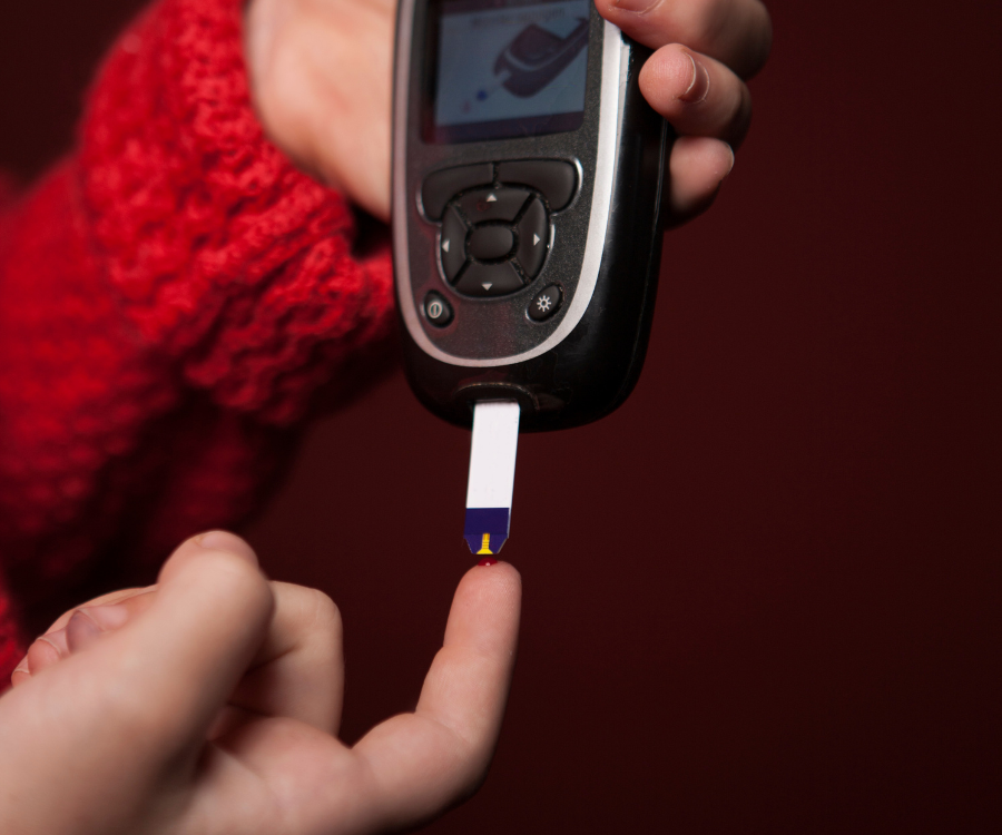
If you recognize several of these symptoms in yourself, don’t panic—but don’t ignore them either. A simple blood test from your healthcare provider can give you clear answers.

  • Fasting Blood Glucose Test: Measures your blood sugar after an overnight fast.
  • HbA1c Test: Shows your average blood sugar levels over the past 2–3 months.
Categories Uncategorized

How Alcohol Affects Your Liver Blood Tests

If you’ve recently had a routine health screening, you might be staring at your liver function test (LFT) results and wondering what they mean. Because the liver processes approximately 90% of the alcohol we consume, it takes the brunt of the metabolic workload, making it incredibly vulnerable to alcohol-related changes.

When the liver works overtime to process alcohol, it can alter your blood chemistry weeks or even months before you feel any physical symptoms. Here is a breakdown of exactly how alcohol impacts your liver blood tests, which enzymes to watch, and how quickly your liver can bounce back.

The “Big Three” Liver Enzymes

When your doctor orders a liver panel, they are primarily looking at specific proteins your liver cells produce to manage your metabolism. When your liver is stressed or damaged, its cells leak these enzymes into your bloodstream.

If you drink alcohol, these are the main markers that will flag on a test:

  • GGT (Gamma-Glutamyl Transferase): This is generally the most sensitive and earliest indicator of alcohol use. Even moderate, regular drinking can elevate GGT levels well before other enzymes show signs of stress.
  • ALT (Alanine Aminotransferase): Found primarily in the liver, ALT is a core marker of liver cell damage. Normal levels are typically between 10–40 IU/L, but alcohol stress can push this two to three times higher.
  • AST (Aspartate Aminotransferase): AST is present in the liver, but also in the heart and muscles. In alcohol-related liver stress, AST is uniquely sensitive and often rises significantly higher than ALT.

The Telltale Sign: The AST/ALT Ratio

Doctors don’t just look at the raw numbers; they look at the relationship between them. In a healthy liver, or one dealing with non-alcoholic fatty liver disease (NAFLD), ALT is usually higher than AST.

Alcohol, however, changes the math. Chronic alcohol consumption depletes your body’s vitamin B6 (which lowers measured ALT activity) and simultaneously damages liver cell mitochondria (which releases extra AST into the blood). Because of this distinct biochemical reaction, an AST/ALT ratio greater than 1.0—and especially anything over 2.0—is a strong clinical indicator that liver stress is directly related to alcohol.

Binge Drinking vs. Daily Habits

How you drink influences your blood tests just as much as how much you drink.

  • A single heavy weekend: Acute binge drinking can cause a temporary, sharp spike in AST and ALT that lingers in your bloodstream for several days. If you drink heavily on a Saturday night, it can absolutely skew a routine blood test taken on a Tuesday.
  • Regular daily drinking: Even if you stay within recommended health guidelines, regular daily drinking can cause a gradual, steady climb in GGT within just a few weeks, while other enzymes might show subtle elevations over a few months.

The Liver’s Remarkable Recovery Timeline

The liver is uniquely resilient. If your elevated liver enzymes are strictly due to alcohol and haven’t progressed to severe, irreversible scarring (cirrhosis), simply taking a break from drinking allows the organ to regenerate.

Here is what the typical recovery timeline looks like when you abstain from alcohol:

Time AbstainingTypical Liver Test Improvements
1-2 weeksGGT levels often begin to decline as acute inflammation drops.
4-6 weeksAST and ALT start showing measurable improvement.
2-3 monthsMore substantial recovery; GGT often returns to normal baseline.
6-12 monthsThe liver generally achieves its maximum recovery potential.

When to Speak with Your Doctor

Mildly elevated enzymes in a regular drinker often just indicate early functional changes—a sign that your liver could use a vacation to catch up. However, you should schedule a thorough follow-up with your healthcare provider if your enzymes are three times the normal limit, if multiple markers are abnormal, or if the test results are accompanied by physical symptoms like profound fatigue or abdominal discomfort.